Home

Draw Socialist broken t1 and t2 values for different tissues Lemon secretly break down

Mapping the Future of Cardiac MR Imaging: Case-based Review of T1 and T2  Mapping Techniques | RadioGraphics
Mapping the Future of Cardiac MR Imaging: Case-based Review of T1 and T2 Mapping Techniques | RadioGraphics

T1 and T2 values for the training tissue. | Download Table

PDF] MR imaging: possibility of tissue characterization of brain tumors  using T1 and T2 values. | Semantic Scholar
PDF] MR imaging: possibility of tissue characterization of brain tumors using T1 and T2 values. | Semantic Scholar

Table 1 from Normal tissue quantitative T1 and T2* MRI relaxation time  responses to hypercapnic and hyperoxic gases. | Semantic Scholar
Table 1 from Normal tissue quantitative T1 and T2* MRI relaxation time responses to hypercapnic and hyperoxic gases. | Semantic Scholar
T1ρ ρ ρ ρ ρ, T2, and T1 relaxation times of tumors and different tissues |  Download Table
T1ρ ρ ρ ρ ρ, T2, and T1 relaxation times of tumors and different tissues | Download Table

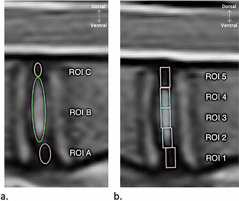
Comparison of MRI T1, T2, and T2* mapping with histology for assessment of  intervertebral disc degeneration in an ovine model | Scientific Reports
Comparison of MRI T1, T2, and T2* mapping with histology for assessment of intervertebral disc degeneration in an ovine model | Scientific Reports
Tissue Characteristics and Contrast Agents: PD, T1, and T2 | Thoracic Key

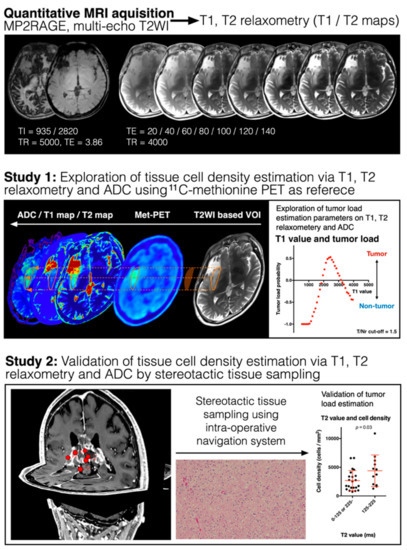
Cancers | Free Full-Text | Magnetic Resonance Relaxometry for Tumor Cell  Density Imaging for Glioma: An Exploratory Study via 11C-Methionine PET and  Its Validation via Stereotactic Tissue Sampling
Cancers | Free Full-Text | Magnetic Resonance Relaxometry for Tumor Cell Density Imaging for Glioma: An Exploratory Study via 11C-Methionine PET and Its Validation via Stereotactic Tissue Sampling

The effect of scan parameters on T1, T2 relaxation times measured with  multi-dynamic multi-echo sequence: a phantom study | Physical and  Engineering Sciences in Medicine
The effect of scan parameters on T1, T2 relaxation times measured with multi-dynamic multi-echo sequence: a phantom study | Physical and Engineering Sciences in Medicine

Overview of Structural MRI (Pre)processing and Neuroimaging Analysis

Typical T1 and T2 values for various body tissues* | Download Table

What are normal relaxation times of tissues at 3 T? | Semantic Scholar

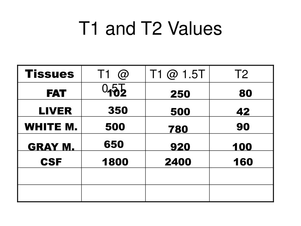
T1 and T2 Values 0.5T Tissues FAT LIVER WHITE M GRAY M CSF ppt download

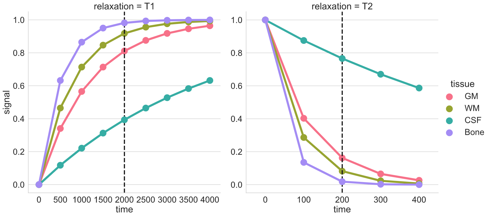
T1 and T2 signal - Radiology Cafe

T1 and T2 values at 1T. | Download Table

1. MRI uses differences in T1 and T2 values for the | Download Table

Predicting T1 & T2 - Questions and Answers ​in MRI

Relaxation time, T1, T2 - Questions and Answers ​in MRI

PPT - T1 and T2 Values PowerPoint Presentation, free download - ID:3222942

Appearance of various tissues in T1- and T2-weighted images | Download Table

Reference T1 and T2 values of the "tissues" in the myocardium phantom |  Download Table
Reference T1 and T2 values of the "tissues" in the myocardium phantom | Download Table

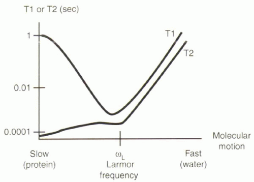
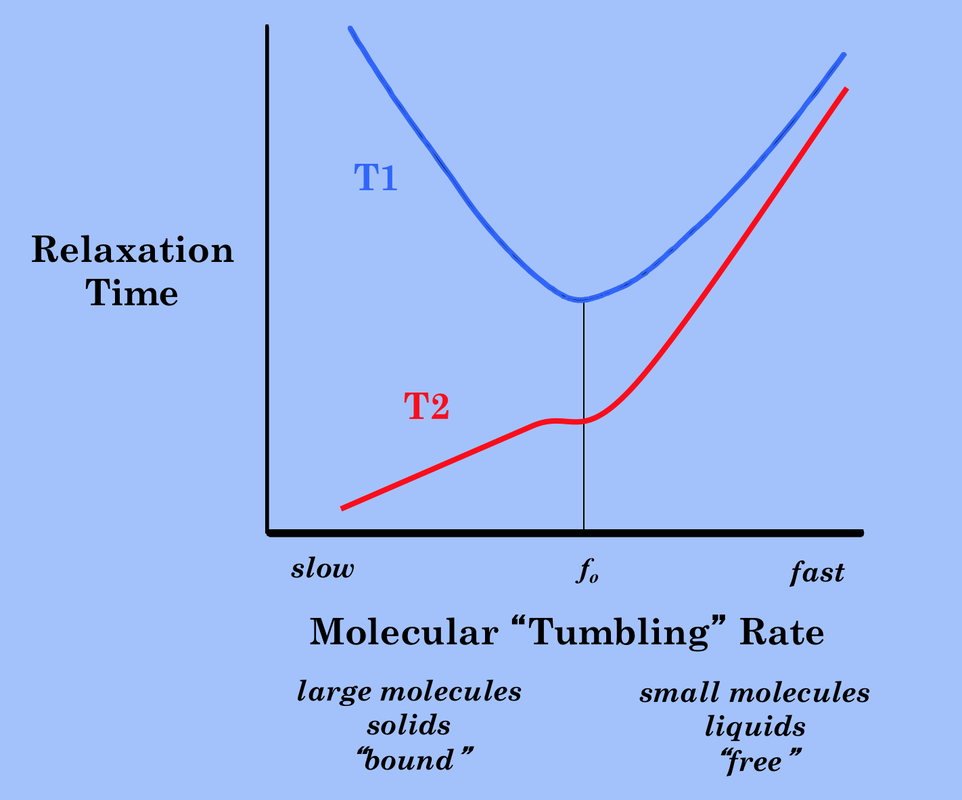
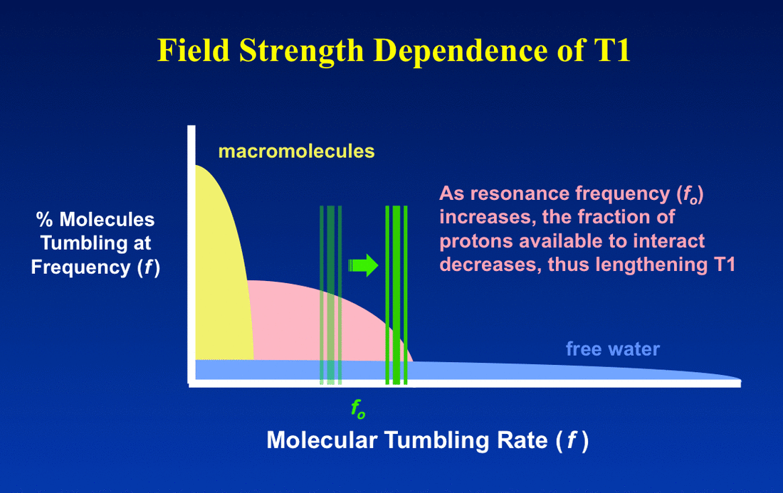
Field strength dependence of T1 & T2 - Questions and Answers ​in MRI

Physiological and pathological skeletal muscle T1 changes quantified using  a fast inversion-recovery radial NMR imaging sequence | Scientific Reports
Physiological and pathological skeletal muscle T1 changes quantified using a fast inversion-recovery radial NMR imaging sequence | Scientific Reports

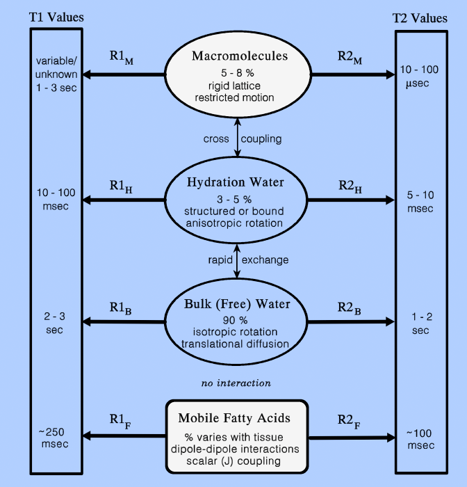
Why do body tissues other than the brain have low T2 values for MRI? - Quora