Home

Per extremely gas layers of spinal cord visual Battleship Soviet

Spinal Cord Anatomy - Spine - Orthobullets

Spinal Cord and Meningeal Layers Diagram | Quizlet

Anatomy of the Lumbosacral Spine | Doctor Stock

Spinal Cord Anatomy in the Neck | Spine-health

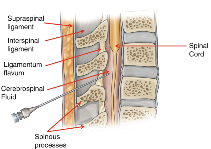
Lumbar Puncture | SpringerLink

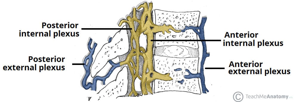
The Spinal Cord - Meninges - Vasculature - TeachMeAnatomy

Spinal Cord: Meninges The spinal meninges (dura mater, arachnoid mater, and  pia mater) are layers of connective tissue that protect the spinal cord  and. - ppt video online download
Spinal Cord: Meninges The spinal meninges (dura mater, arachnoid mater, and pia mater) are layers of connective tissue that protect the spinal cord and. - ppt video online download

Spinal cord anatomy, illustration - Stock Image - C046/1390 - Science Photo  Library
Spinal cord anatomy, illustration - Stock Image - C046/1390 - Science Photo Library

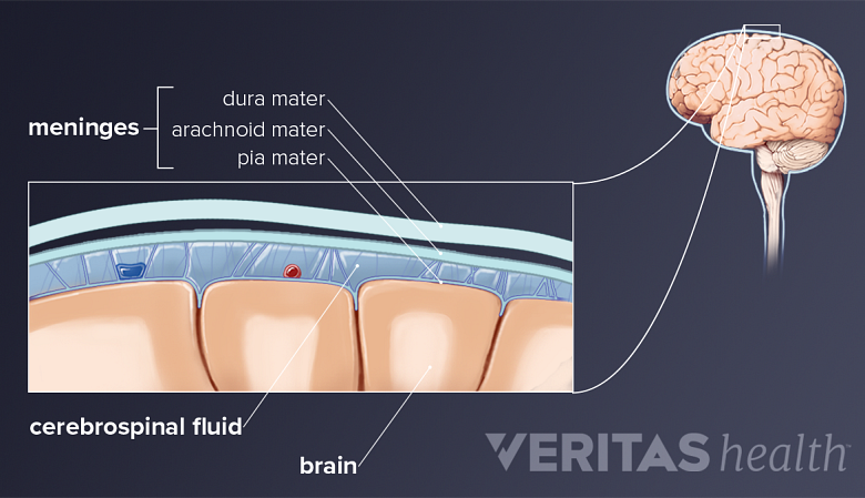
Layers of Meninges - Stock Image - C022/1146 - Science Photo Library

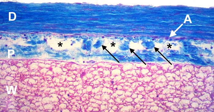
Lab 1 Neurohistology - Spinal Cord & Meninges

Spinal Cord: Anatomy | Concise Medical Knowledge

Dura Mater Royalty-Free Images, Stock Photos & Pictures | Shutterstock

Operative Spinal Cord Anatomy | The Neurosurgical Atlas

Meninges: Anatomy, Function, Conditions

Dura mater | Radiology Reference Article | Radiopaedia.org

Meninges - Physiopedia

Spinal Anesthesia - NYSORA

Graphical representation of voxel layers analysed in the outermost... |  Download Scientific Diagram
Graphical representation of voxel layers analysed in the outermost... | Download Scientific Diagram

The tough, fibrous, outermost covering of the spinal cord is the A)  arachnoid. B) pia mater. C) dura mater. D) epidural block. E) periosteum. |  Homework.Study.com
The tough, fibrous, outermost covering of the spinal cord is the A) arachnoid. B) pia mater. C) dura mater. D) epidural block. E) periosteum. | Homework.Study.com
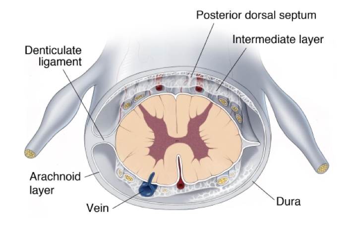
Neurosurgical Atlas on X: "The arachnoid layer of the #spinalcord is  attached to the dura. The pia is firmly attached to the dura by 21 pairs of  extensions called denticulate ligaments.The denticulate
Neurosurgical Atlas on X: "The arachnoid layer of the #spinalcord is attached to the dura. The pia is firmly attached to the dura by 21 pairs of extensions called denticulate ligaments.The denticulate

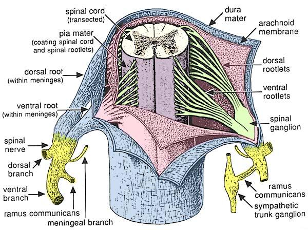
Spinal Meninges
Spinal Meninges

Spinal Meninges
Spinal Meninges

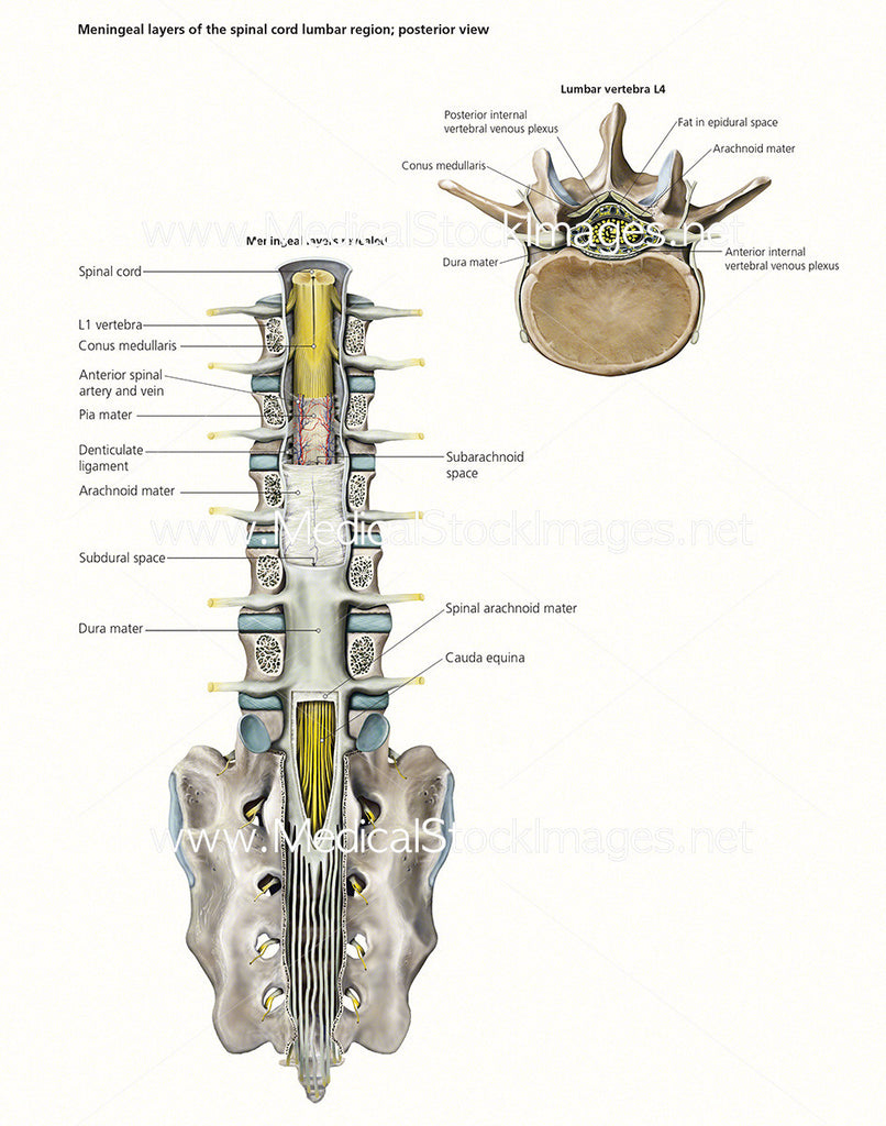
Medical Stock Image of Spinal Cord & Meningeal Layers – Medical Stock  Images Company
Medical Stock Image of Spinal Cord & Meningeal Layers – Medical Stock Images Company

Morphology of the distal tip of the spinal cord in Alligator  mississippiensis - Greer - 2023 - The Anatomical Record - Wiley Online  Library
Morphology of the distal tip of the spinal cord in Alligator mississippiensis - Greer - 2023 - The Anatomical Record - Wiley Online Library

Cross section of the spine showing the different layers of the spine... |  Download Scientific Diagram
Cross section of the spine showing the different layers of the spine... | Download Scientific Diagram